Magnetic resonance imaging (MRI), is a scanning technique for creating detailed images of the human body.
The scan uses a strong magnetic field and radio waves to generate images of parts of the body that can’t be seen as well with X-rays, CT scans or ultrasound. For example, it can help doctors to see inside joints, cartilage, ligaments, muscles and tendons, which makes it helpful for detecting various sports injuries.
MRI is also used to examine internal body structures and diagnose a variety of disorders, such as strokes, tumors, aneurysms, spinal cord injuries, multiple sclerosis and eye or inner ear problems.
What to expect
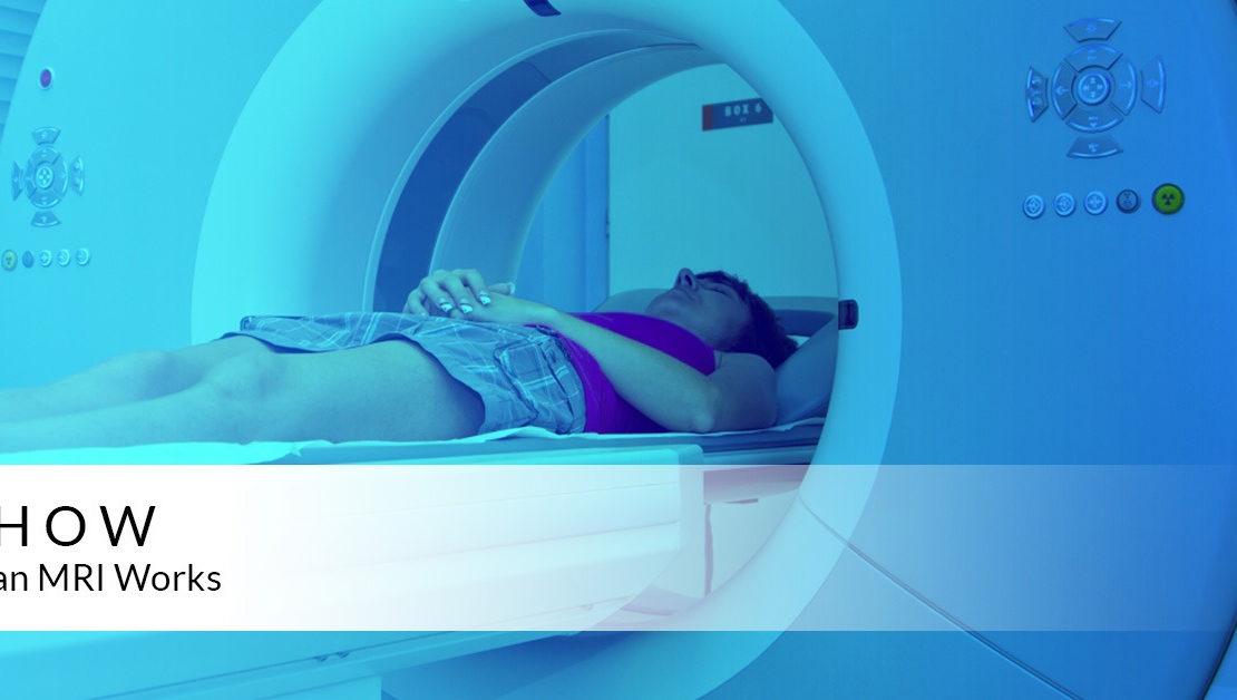
During an MRI, a person will be asked to lie on a movable table that will slide into a doughnut-shaped opening of the machine to scan a specific portion of your body. The machine itself will generate a strong magnetic field around the person and radio waves will be directed at the body.
A person will not feel the magnetic field or radio waves, so the procedure itself is painless. However, there may be a lot of loud thumping or tapping noises during the scan, so people are often given headphones to listen to music or earplugs to help block the sound. At MRI Unit ( Institute Of Child Health ), we got the most silent MRI machine from Siemens.
Some people may be given a contrast solution by intravenous, a liquid dye that can highlight specific problems that might not show up otherwise on the scan.
Young children as well as people who feel claustrophobic in enclosed places may be given sedating medication to help them relax or fall asleep during the scan because it is important to stay as still as possible to get clear images. Movement can blur the images. At MRI Unit ( Institute Of Child Health ), we got the wide bore MRI machine from Siemens, which significantly reduces the chances of claustrophobia.
How it works
The human body is mostly water. Water molecules (H2O) contain hydrogen nuclei (protons), which become aligned in a magnetic field. An MRI scanner applies a very strong magnetic field (about 0.2 to 3 teslas, or roughly a thousand times the strength of a typical fridge magnet), which aligns the proton “spins.”
The scanner also produces a radio frequency current that creates a varying magnetic field. The protons absorb the energy from the magnetic field and flip their spins. When the field is turned off, the protons gradually return to their normal spin. The return process produces a radio signal that can be measured by receivers in the scanner and made into an image. Protons in different body tissues return to their normal spins at different rates, so the scanner can distinguish among various types of tissue. A radiologist interpret these images to clinical findings.